Introduction

In the world of medical diagnostics, the ability to detect diseases accurately and swiftly is paramount. In this data science project, I set out to explore the world of deep learning to detect brain tumors from medical images. With a dataset of 253 brain images, consisting of 155 cases with brain tumors and 98 cases without, I aimed to build a predictive model using the powerful VGG16 architecture. This blog post will take you through the journey of this project, from data preprocessing to model training and evaluation.
Dataset
Gathering and Preparing the Data
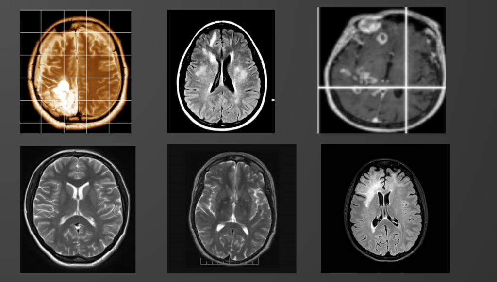
The dataset I used in this project was a collection of brain MRI images. It consisted of 253 images, of which 155 were cases of brain tumors, and 98 were healthy brain images. The dataset was carefully curated and labeled, making it an ideal starting point for our brain tumor detection task.
Data Preprocessing

Before feeding the images into the model, several preprocessing steps were performed:
Image Resizing: All images were resized to a uniform size (e.g., 224×224 pixels) to ensure consistency for model training.
Normalization: Image pixel values were scaled to the range [0, 1] to facilitate convergence during training.
Data Augmentation: To enhance the model’s ability to generalize, data augmentation techniques such as rotation, flipping, and zooming were applied to create additional training samples.
Building the Model
For this project, I chose to implement the VGG16 model, a deep convolutional neural network architecture known for its effectiveness in image classification tasks. VGG16 consists of multiple convolutional and max-pooling layers followed by fully connected layers.
Training and Evaluation
With the model architecture in place, it was time to train and evaluate its performance. The dataset was split into training and testing sets to assess the model’s ability to generalize to unseen data.
Model Training
The model was trained using the training set with binary cross-entropy loss and the Adam optimizer. Training was carried out for a specified number of epochs, monitoring both training and validation accuracy.
Model Evaluation
The trained model’s performance was evaluated using the testing set. Key evaluation metrics included accuracy, precision, recall, and F1-score. These metrics provided insights into the model’s ability to correctly classify brain tumor images.
Results
After training and evaluation, the model achieved an accuracy of approximately 82%. This meant that the model correctly classified brain tumor images 82% of the time. Furthermore, the model’s precision, recall, and F1-score indicated a good balance between true positives and false positives.
Conclusion
In this data science project, we leveraged deep learning and the VGG16 architecture to detect brain tumors from medical images. With careful data preprocessing, model building, and evaluation, we achieved an accuracy of 82%, showcasing the potential of deep learning in medical diagnostics.
While this project is a promising step forward, there is still room for improvement. Future work could involve fine-tuning hyperparameters, exploring different neural network architectures, and expanding the dataset to enhance model performance and robustness.
Brain tumor detection using deep learning holds immense promise for early diagnosis and improved patient outcomes, and this project serves as a testament to the power of data science in healthcare.
Thank you for joining me on this journey into the world of brain tumor detection with deep learning. Stay tuned for more exciting data science adventures!

